
Reading time: 7 minutes
Michael Marand
In short-distance track competitions, the winner of a race is largely determined by how well the runners take off from the starting blocks. With the athletes only narrowly separated by their top speeds, the initial difficulty of shifting from an immobile crouch to rapid acceleration is an art that distinguishes the victor from his or her competitors. Meanwhile, in cancer care, surgery, chemotherapy, and radiotherapy are powerful tools in treating many different cancers. Yet, cancers are elusive, persistent, and heterogeneous, often leading to difficulty achieving overall treatment success. It follows that patient outcomes are often influenced by how much of a head start physicians can get on treating the cancer.
However, several factors can lead to delays in cancer patient treatment. First, a patient with unknown complications may be hesitant to seek diagnostic attention due to unease about procedures or financial concerns. Similarly, a primary care provider may spend time attempting to rule out other conditions before referring a patient for a more invasive diagnostic procedure. Additionally, even after the test is administered, it can take a few weeks for the test results to be finalized. A rapid, non-invasive, and cost-effective cancer screening tool could provide value to patients by allowing them to undergo cancer intervention sooner.
In this theme, a team from Terasaki Institute recently shared its progress towards engineering contact lenses to detect cancer. The tool relies on naturally occurring messenger molecules called exosomes. Exosomes encase and transport protein, genetic material, and lipids from one cell to another. These exosomes are approximately a thousand times smaller than most human cells. Exosomes are an intriguing target for cancer diagnosis because they are constructed within the originating cell before they are released for transport. Consequently, the composition of an exosome gives clues about the state of the cell it originated from. Specifically, the proteins on the surface of an exosome are related to those of the originating cell. The Terasaki Institute team intends to take advantage of the elevated presence of specific proteins, such as growth factors, on cancer cell exosomes. The presence of this kind of exosome is a signal that cancer may be present somewhere in the body. The researchers chose to search for these exosomes in tears. Advantageously, tears do not contain much of the cellular debris that is present in other fluids such as blood, urine, and saliva.
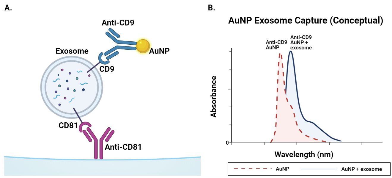
The first step in achieving this aim was engineering contact lenses that could physically capture exosomes. The mechanism that was used to capture exosomes in this study relies on antibodies. Antibodies are proteins produced by immune cells that bind to specific counterpart proteins called antigens. The team chemically attached anti-CD81 antibodies to the surface of contact lenses. These anti-CD81 antibodies bind exclusively to the CD81 antigen on the surface of exosomes. The antibodies were placed within laser-engraved microchambers in the contact lens to prevent them from rubbing off on contact with the eye. Cleverly, the microchambers were beveled inwards to guide tears into the cavity housing the antibodies. To assess whether exosomes were captured, a common analytical technique called UV-vis spectroscopy was employed. UV-vis spectroscopy works by exposing a sample to different wavelengths of light and measuring the amount of light transmitted through the sample. With these records, one can easily produce a graph of the amount of light absorbed by the sample at each wavelength tested. The wavelength at which the absorption of light is maximized is a useful metric for comparing samples. In the present case, anti-CD9 antibodies bound to gold nanoparticles bind CD9 antigens on the surface of exosomes. On their own, these anti-CD9 gold nanoparticles absorb light most strongly at 549 nm. When attached to a protein, such as CD9 antigen on the exosome, the wavelength of maximum absorbance increases to about 557 nm. Therefore, the amount of 557 nm light absorbed can be used to quantify the success of exosome capture by an engineered lens sample.
These engineered contact lenses should ideally be capable of detecting exosomes from a broad range of cancer types. In this theme, an early experiment assessed if the lenses could detect exosomes from 10 different cell lines covering a range of different physiological tissues. Each different cell line was grown in a separate flask with liquid supporting cell growth. Liquid was drawn from each flask and incubated with the anti-CD81 lenses. The lens surface was washed and then dosed with anti-CD9 gold nanoparticles. For each sample, the amount of absorption at 557 nm was significantly greater than the amount of absorption of the negative control sample, which contained cell growth liquid that had not been in contact with cells. Therefore, exosomes were indeed captured from all 10 different cell lines.
The next step was to validate the ability of the engineered lenses to distinguish between cancerous and normal exosomes. In this experiment, 3 cell lines were chosen. MCF 10A is a non-cancerous mammary gland cell line that does not express HER2 or ER receptor proteins. HER2 and ER are receptor proteins that are associated with cancer cell division. MCF 7 is a cancerous breast cancer cell line that expresses both HER2 and ER. MDAMB231 is metastatic breast cancer that produces a small amount of HER2, but not ER. Just as it was done in the previous experiment, liquid was taken from each flask and incubated with the anti-CD81 lenses. However, this time several different gold nanoparticles were added. In addition to the anti-CD9 gold nanoparticles, particles conjugated to anti-HER2 and anti-ER antibodies were added. As expected, all 3 cell lines reported CD9 expression, confirming that exosomes in general were present. The non-cancerous MCF 10A was indeed negative for both HER2 and ER. As for the cancerous cell lines, in alignment with previously reported expression profiles, MCF7 was positive for both HER2 and ER while MDAMB 231 was positive for HER2 only. These results indicate that the engineered contact lenses can discriminate between exosomes from cancerous and non-cancerous cell lines if supplied with the appropriate antibodies.
As the experiments described thus far were conducted in cell growth liquid, the team then investigated if the lenses work in real tears and if they are safe for the eye. In this phase, the lenses were tested in tear samples from 10 different volunteers. Encouragingly, exosomes were successfully detected in all tear samples. To assess biocompatibility, the engineered contact lenses were incubated with mouse connective tissue cells. After 7 days, the team found no significant change in the viability of these cells.
In this study, the authors demonstrate that a contact lens engineered with antibody-containing microchannels can detect exosomes from a variety of cancerous and healthy cells. A great success herein is the detection of exosomes without needing to undertake the time-consuming process of isolating the exosomes. In the short term, the authors plan to validate their findings in animal models. The authors also claim that with some modifications these engineered lenses could have other applications beyond cancer diagnostics. For example, the lenses may have utility in diagnosing other diseases and could even be used to deliver drugs.
While these lenses will not replace traditional diagnostics, they have the potential to be a valuable supplemental option. The lenses could provide a rapid, cost-effective early step in a patient’s cancer screening that ultimately reduces the amount of time it takes for a patient to begin treatment. Further validation and optimization are requisite, but this Terasaki Institute team has made a phenomenal jump out of the starting blocks with this initial study.
Edited by Emily Chan
Works Discussed:
Li S, Zhu Y, Haghniaz R, et al. A microchambers containing contact lens for the noninvasive detection of tear exosomes. Advanced Functional Materials. 2022:2206620. doi:10.1002/adfm.202206620
Tsai W-C, Kung P-T, Wang Y-H, Kuo W-Y, Li Y-H. Influence of the time interval from diagnosis to treatment on survival for early-stage liver cancer. PLOS ONE. 2018;13(6). doi:10.1371/journal.pone.0199532

Leave a comment