
Darshak Bhatt
Reading time: 4 minutes
Migration is tough! The International committee for the Red Cross states that “… on their journey, migrants face multiple risks and high degree of vulnerability”, and adds “Thousands (of migrants) die or disappear along the way every year.” Similarly, a cancer cell faces challenges related to migration and the seemingly impossible task of survival during the process of metastasis, the spread of cancer through the body. Upon reaching such distant sites, cancer cells can seed and form secondary tumors called metastases. Needless to say, there are some factors that shape the success of these cancer cells before they even leave the tumor in the first place.
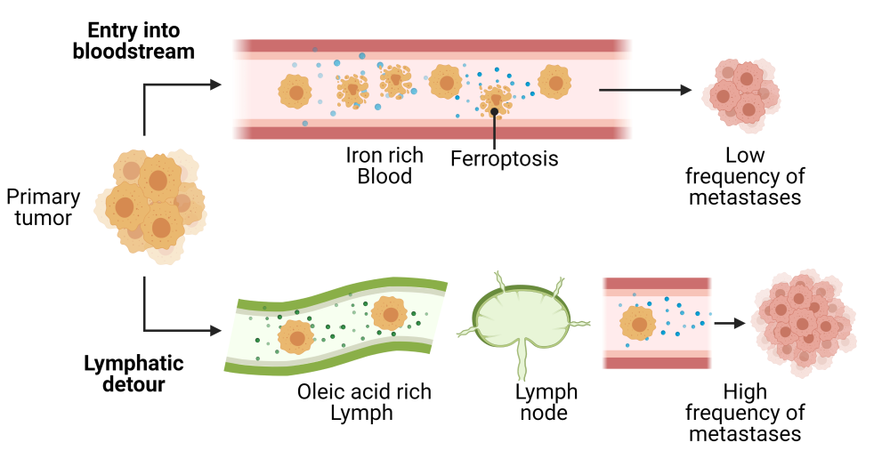
The connecting link or the ‘highway’ between a tumor and distant organs is often the vascular system, consisting of the lymphatic system and the circulatory system. The lymphatic circuit differs from the blood circuit in composition (lymph is richer in fatty acids, does not contain red blood cells, has fewer immune cells, and lacks plasma proteins), flow-rate (lymph is slower than blood), and various other physiological functions. Interestingly, it has been reported that skin cancer cells take a regional lymphatic system detour before metastasizing systematically through blood circulation . A recent study explains that these cancer cells undergo less stressful experiences in the lymph than in the blood, which improves their chances of migration and successful metastasis. Amidst a variety of stress exerted by the blood environment, the researchers found that iron-dependent oxidative stress called ferroptosis plays a major role in causing cancer cell death during migration and thus limits the possibility of forming metastasis. Ferroptosis occurs via accumulation of lipid-based oxidant molecules inside cancer cells causing damage to biomolecules and an imbalance in cellular homeostasis. On the other hand, melanoma cells that had a lymphatic detour prior to being exposed to the stressful blood environment were better prepared to resist such oxidative stress.
(Adapted from Nature news and views)
Exploring this idea, the researchers injected melanoma cells into the skin of mice with or without a fully functional immune system, and found that there was a higher frequency of melanoma cells in the lymph nodes associated with tumors compared to the tumor associated blood vessels. By measuring cellular oxidative stress, the researchers found that melanoma cells consistently exhibited higher levels of oxidative stress in the blood than in the primary tumors and lymph. To define the causative agent of oxidative stress in cancer cells, they treated melanoma cells with various inhibitors of oxidative stress caused by different molecular pathways and found that only via inhibition of ferroptosis (an iron-dependent form of cell death) was there an increase in the survival and metastatic potential of cancer cells via blood.
Next, the researchers started comparing features between blood and lymph that may play a key role in regulating ferroptosis-induced cancer cell death. They found that higher levels of free iron and lower levels of oleic acid in the blood contribute to cancer cell death. From previous studies, we have learned that cells can acquire free iron from their environment, which stimulates ferroptosis. Alternatively, the enriched presence of mono-unsaturated lipid molecules like oleic acids can inhibit ferroptosis. Moreover, consistent with their findings, the researchers found that melanoma cells pretreated with oleic acid survived better in the blood after intravenous injection into mice as compared to untreated melanoma cells. Additionally, if iron was removed from the media surrounding melanoma cells, ferroptosis could not occur.
Finally, the authors investigated whether lymph node metastasis occurs before all other locations and whether cancer cells are primed to withstand ferroptosis in the lymph, leading to successful spread. To this end, they collected mouse melanoma cells from skin tumors and from lymph-node tumors, and injected these cells into the blood circulation. They found that melanoma cells from lymph nodes had better chances to form secondary tumors as compared to cells found in skin tumors. Additionally, cancer cells from the skin tumors were more sensitive to treatment with the ferroptosis-inducing molecule erastin than the cells from lymph-node tumors.
These results suggest that a little help from the lymphatic environment allows cancer cells to incorporate oleic acid and other antioxidants that later protect the cells from ferroptosis during the stressful route through the blood before reaching those distant sites of new settlements.
Edited by Sara Musetti
Work Discussed:
Ubellacker, J.M., Tasdogan, A., Ramesh, V. et al. Lymph protects metastasizing melanoma cells from ferroptosis. Nature 585, 113–118 (2020). https://doi.org/10.1038/s41586-020-2623-z
Illustrations: Made in Bio-Render.
Darshak Bhatt is a PhD student at the department of Experimental Oncology at University of São Paulo and at university medical center Groningen. You can find him at his blog or on twitter @DarshakWrites.
If you are curious about the role of lymphatic system and ferroptosis in cancer, read more here:
- How lymphatic system intertwines with tumor spread: https://oncobites.blog/2020/05/13/building-a-highway-to-tumors/
- How tumor draining lymph nodes help spread tumor: https://oncobites.blog/2018/04/25/in-cancer-your-own-lymph-nodes-turn-against-you/
- How ferroptosis leads to cancer cell death: https://oncobites.blog/2020/04/22/ferocious-ferroptosis-programmed-cell-death-in-cancer-treatment/
- Red cross statement on challenges of migration: https://www.icrc.org/en/document/speech-migration-and-internal-displacement-national-and-global-challenges
