
Sara Musetti
Estimated reading time: 5 minutes
It seems talking about the immune system and how it can fight cancer is all the rage these days, especially here at OncoBites. But talking about it can be tricky, especially because most people seldom think about their immune system. It’s a part of your body, but… which part? It’s not like an arm, or a heart, that you can pinpoint. So, where do these elusive immune cells and their alleged cancer fighting powers reside? The answer is your lymphatic system!
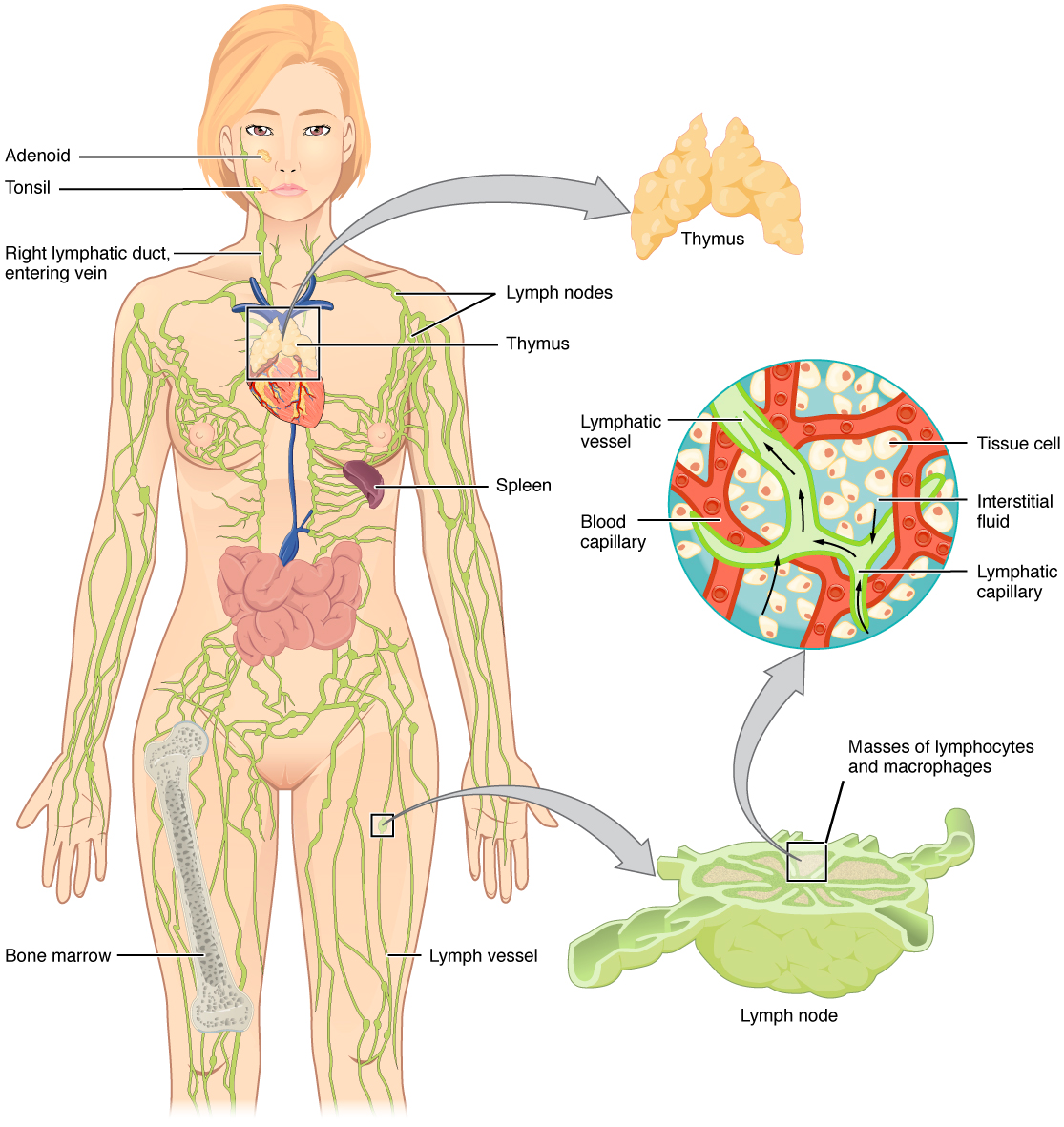
As a mirror to the circulatory system, the lymphatic system is a network of vessels that connects our immune organs, most notably the thymus and the spleen. Spread throughout our lymphatic system are lymph nodes, specialized areas where immune cells (like the B cells and T cells) reside and scan the lymphatic fluid from nearby tissue for signs of infection, illness or damage. While most people only notice their lymph nodes when they suddenly balloon-up during illness, the lymphatic system is of critical importance for our overall health. When immune cells are activated, they multiply and move from the lymph node into the surrounding tissue where they detected the threat. That is why a cold makes our lymph nodes swell—it’s all the immune cells getting ready to come to our rescue!
In cancer research, lymph nodes can be complicated. Because lymph flows from tissues directly to lymph nodes, lymph nodes are often one of the first sites of metastasis. Dr. Emily Harrison wrote about this fear in 2018 in one of our earliest articles. But, as we know, using the immune system to fight cancer is increasingly popular, which begs the question: Can strengthening the lymphatic system actually be used to fight cancer?
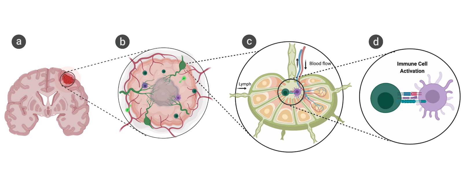
To answer this problem, cancer immunologists at Yale Medical School led by Dr. Akiko Iwasaki decided to tackle one of the most difficult cancers head-on: they were going to boost lymphatic drainage in glioblastoma, a deadly form of brain cancer. To do this, they used a virus known to target the brain but cause no damage on its own, called adeno-associated virus 9 (AAV-9), to carry a gene for a protein that enhances vessel growth, called VEGF-C. They tested two different models: In one, they treated mice with glioblastoma with the AAV9-VEGF-C and observed that tumors were almost completely eradicated. In the second, they gave mice the VEGF-C virus after they had removed lymph nodes that receive lymph from the brain, then they implanted the mouse brains with glioblastoma cells. In this way, they could study whether having more lymphatic drainage helped reduce the growth of brain cancer without getting the immune system involved, because removing the lymph nodes removes the ability of the immune system to check the brain for disease. While the tumors in mice with VEGF-C treatment did grow slower than those without treatment, the lack of lymph nodes meant that the tumors did still grow. However, if lymph nodes were left intact and mice were treated with VEGF-C, tumors shrank and the mice survived. Dr. Iwasaki’s team was able to link the administration of AAV9-VEGF-C specifically to the formation of more lymph vessels in the brain, not the formation of blood vessels, and to link these new lymphatic vessels with anti cancer immunity.
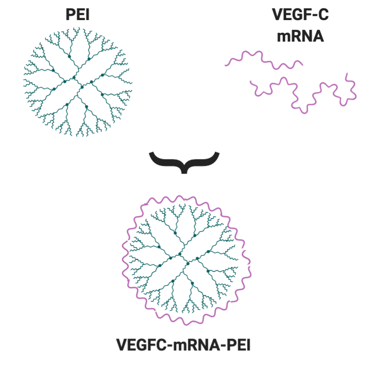
To confirm the role of the immune system in this therapy and demonstrate the value of this work in the clinic, the team moved to using a method of VEGF-C delivery that would be more useful for treating patients. They used a chemical known as PEI to hold mRNA that tells cells to produce VEGF-C and carry it to the cells in the brain that can make more lymphatic vessels. Picture PEI almost like an octopus, with lots of tentacles and suction cups for grabbing things. Instead of tentacles, PEI has branches, each of which has lots of sites to “grab” mRNA. PEI has lots of positive groups, and mRNA is negative, so they stick together very well. When PEI and mRNA are mixed, they make very small bundles that can be safely injected into patients. mRNA cannot be delivered alone because it’s too delicate and the body degrades free-floating mRNA in case it comes from a virus, so bundling it up with PEI helps protect it.
Once again, VEGF-C, this time carried by PEI-mRNA complexes, was able to protect mice from glioblastoma and from other cancers in the brain, including melanoma. In addition, the new lymphatic vessels in the brain were able to improve the efficacy of popular checkpoint inhibitors in brain cancer. Checkpoint inhibitors work by helping release the brakes on the immune system so it can kill tumor cells, and because the increase in the lymphatic system in the brain helps increase immune cell access to the tumor, VEGF-C delivery boosts checkpoint inhibitor therapy. This could be a groundbreaking shift for brain cancer, as checkpoint inhibitors have revolutionized treatment for many cancers and could even help prevent relapse, which is almost always deadly in the case of glioblastoma.
As exciting as this research is, we’re still a long way from understanding the full implications of inducing lymphatic growth in human brains and whether that might be of therapeutic value. While mice are the best model we have for basic research, particularly when we need to study how a tumor interacts with the immune system, we’ve known how to cure cancer in mice for years–those same treatments just don’t always work for humans, or at least not as well. Gene therapies, like delivering VEGF-C mRNA, are also subject to a great deal of scrutiny before they can be applied to human patients, so it is unlikely for a therapy like this to be available soon. However, knowing that VEGF-C can make such a huge difference and that more lymph vessels can mean a strong immune response are both very valuable to the field of cancer research, and I am looking forward to seeing what arises from this work.
Edited by Adil Muneer
Work Cited
Song, E., Mao, T., Dong, H., Boisserand, L.S.B., Antila, S., Bosenberg, M., Alitalo, K., Thomas, J.L. and Iwasaki, A. (2020). VEGF-C-driven lymphatic drainage enables immunosurveillance of brain tumours. Nature, 1-6.
