Reading time: 4 minutes
Cameron Van Cleave
Introduction
If you haven’t heard of amyloid light chain (AL) amyloidosis, you aren’t alone. According to the Amyloidosis Foundation, 4,500 new cases are diagnosed in the US every year. That’s about 0.001% of the population, or roughly 1 in every 10,000 people. That estimate may be low due to misdiagnoses, as AL amyloidosis shares symptoms with a variety of other diseases. This article aims to provide a general overview for the average person to increase awareness.
Understanding AL amyloidosis requires some basic concepts in immunology, namely, the types of immune cells and antibodies. Within the bone marrow are hematopoietic stem cells, which may transform into a variety of different cell types, including red blood cells, white blood cells, and immune cells, as shown in Figure 1. In the context of AL amyloidosis, B cells and plasma cells are key. B cells are immune cells that may be activated upon contact with an antigen, which transforms them into plasma cells.
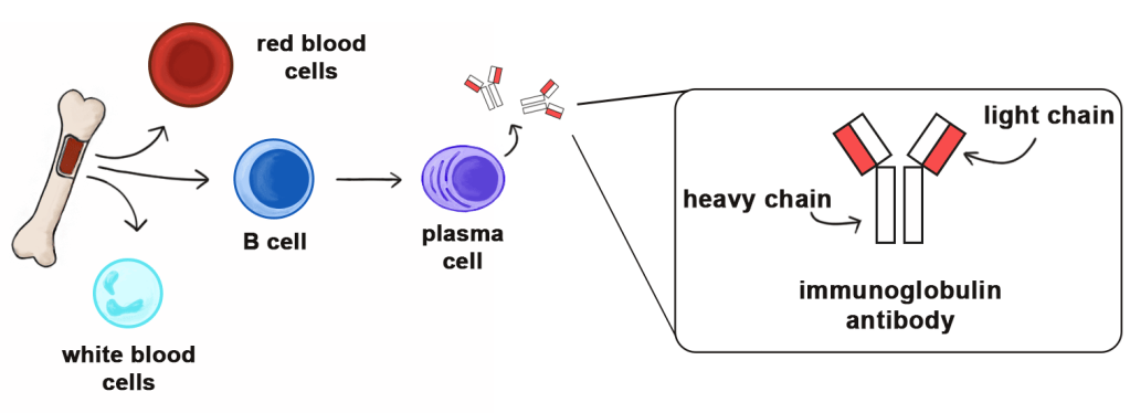
Plasma cells are specialized to produce copious amounts of immunoglobulins, often abbreviated as Ig or Ab. These Igs consist of two heavy chains and two light chains of either λ or κ type. While plasma cells generally secrete full Igs, they sometimes secrete free light chains (FLC), or light chains that are not attached to a heavy chain. These FLCs are the focal point of AL amyloidosis diagnosis and treatment, as they form fibrils that then accumulate in organs.
How the Disease Works
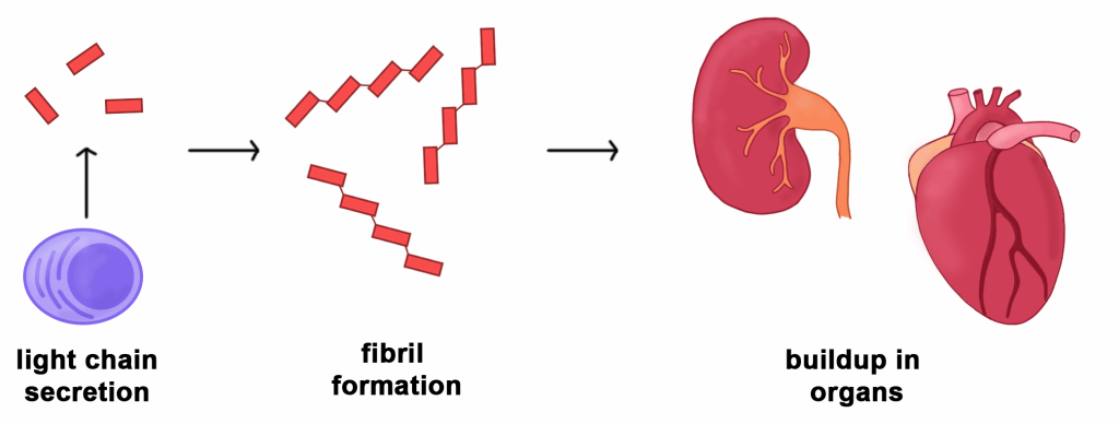
At its core, AL amyloidosis is a disease with a straightforward progression. As shown in Figure 2, a malignant plasma cell in the bone marrow repeatedly replicates itself. These copies secrete misfolded FLCs, which form fibrils. Those fibrils then accumulate in the patient’s organs, leading to organ damage, primarily affecting the heart and kidneys.
Risk Factors
There is no definitive list of risk factors; however, there are clinical overlaps between multiple myeloma and its precursor, MGUS. AL amyloidosis is often co-diagnosed in 10-15% of patients with multiple myeloma and in 1% of patients with MGUS.
Symptoms
The symptoms of AL amyloidosis are nonspecific and differ between patients, making diagnosis difficult. A patient may experience any combination of the following:
- Kidney Issues
- Swelling in the lower legs
- Excess protein in urine
- Kidney failure
- Heart Issues
- Irregular heartbeat
- Shortness of breath
- Gastrointestinal issues
- Nausea
- Chronic fatigue
- Weakness
- Bruising around the eyes
- Swelling of the tongue
Diagnosis
Clinicians use a combination of techniques to diagnose AL amyloidosis:
- Urine tests – High levels of protein in urine may indicate AL amyloidosis and/or kidney issues. Further immunofixation testing may be used to determine if the excess protein is FLC
- Blood tests – A serum FLC assay can quantify FLC levels and determine if the misfolded light chains are λ or κ type
- Bone marrow biopsy – Bone marrow samples are used to determine the relative abundance of abnormal plasma cells, light chain type, and any relevant genetic mutations
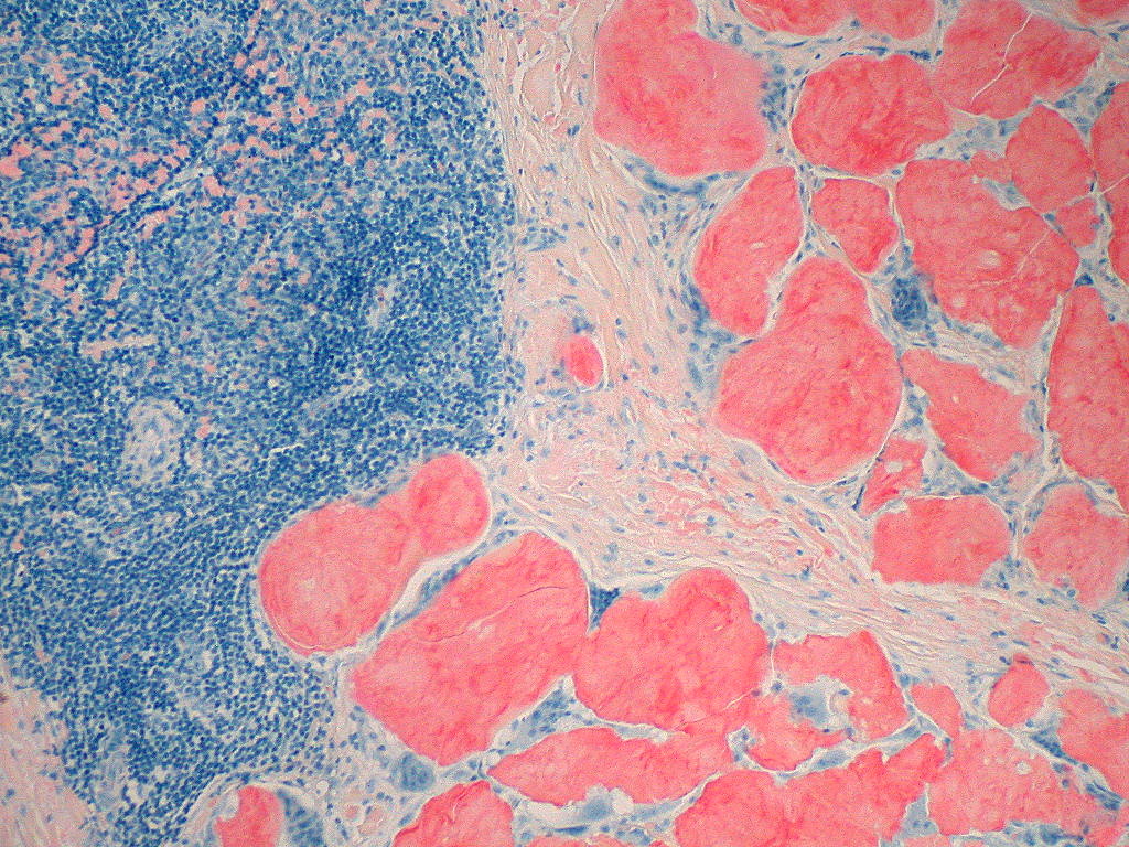
- Fat pad biopsy – A small amount of subcutaneous fat may be smeared on a slide and stained with Congo red. Upon binding amyloid fibrils, Congo red becomes fluorescent, allowing pathologists to view fibrils under a microscope
- Heart tests– Electrocardiograms are used to determine if the heart has been enlarged or stiffened due to the buildup of amyloid fibrils
Treatment
Newly diagnosed patients are assessed to determine their eligibility to receive a peripheral blood stem cell transplant combined with high doses of melphalan. Ideally, this method kills the abnormal plasma cells and allows healthy stem cells to replace them. Approximately 40% of patients who receive a transplant achieve a complete response, meaning their FLC levels in blood and urine return to normal and stay there. Unfortunately, 10-20% of newly diagnosed patients are not eligible for a transplant due to organ dysfunction.
Patients who are ineligible for transplant typically receive a chemotherapy combination known as Dara-CyBorD, which includes daratumamab, cyclophosphamide, bortezomib, and dexamethasone. Most patients (78%) have a good response. This combination may be adjusted depending on genetic mutations, immune status, and the presence of any other medical conditions.
There are currently no established criteria for treating patients who don’t respond to initial Dara-CyBorD therapy or who have relapsed. Doctors may try T cell therapy or prescribe alternative chemotherapy regimens. Still, consideration must be given to genetic mutations, previous therapies, patient fitness, and the extent of organ damage.
Conclusions
AL amyloidosis is a tricky disease to diagnose. The overlap of symptoms with other diseases and patient-specific presentation often leads to misdiagnosis and, therefore, a delay in appropriate treatment. Hopefully, public awareness and education lead more people to recognize the signs and advocate for appropriate care.
Header Image Source: from Flickr.com, Credit: Ed Uthman (image is under a Creative Commons license)
Figure 1 and 2 Image Credits: Created by Author with Adobe Photoshop
Edited by Joycelyn Ghansah, MA, MPH
References
Amyloidosis Foundation. (n.d.). AL Amyloidosis. Retrieved February 14, 2026, from https://amyloidosis.org/al/
Sanchorawala, V. (2024). Systemic Light Chain Amyloidosis. New England Journal of Medicine, 390(24), 2295–2307. DOI: 10.1056/nejmra2304088
Schirrmacher, V. (2023). Bone Marrow: The Central Immune System. Immuno, 3, 289–329. DOI: 10.3390/immuno3030019
 OncoBites
Leave a comment