Morgan McSweeney
A group of researchers from the University of California – San Francisco recently found that the presence of a certain group of immune cells in tumors (“stimulatory dendritic cells,” or SDCs) can predict better cancer outcomes, at least in melanoma patients. For example, in patients treated with checkpoint inhibitors (drugs that work by giving your immune system the “go” signal, allowing it to attack the tumor), more SDCs means better treatment results. These studies suggest that the power of checkpoint inhibitor therapies is much greater when there are greater numbers of natural killer (NK) cells, which are responsible for recruiting the SDCs to the tumor site.
Natural killer cells have two commonly thought-of roles as part of your immune system. They monitor the body for signs of cells infected by viruses, and they keep a watch for early signals that cells might be cancerous. When they find virus-infected cells or cancer cells, they excrete toxins to kill the target cells. They are part of the “innate” immune system, which means that they respond to a broad range of situations, unlike the “adaptive” immune cells, which have been trained to attack one very specific target (such as a bacterial, viral, or cancer-specific protein). NK cells can work together with T cells (which are part of the adaptive immune response) to selectively control cells with cancerous mutations. In the context of these studies, however, NK cells were important because they were playing a different role: they were influencing whether SDCs were attracted to the tumor environment.
This group’s prior research suggested that responses to checkpoint inhibitor therapy were relatively poor in mice that had low numbers of SDCs in their tumors. They found that, without SDCs, the newly-activated T cells were less able to kill cancer cells. The group then analyzed data from human patients and found that fewer SDCs meant worse outcomes. However, although this research was reported in 2014, it has been difficult to translate into clinical action. For one, it can be difficult to assess tissue samples for the presence of SDCs; it would be significantly more convenient and less expensive if there was a blood test that could predict whether tumors would have high or low numbers of SDCs. Further, it hasn’t been understood why some tumors seem to cause the recruitment of SDCs but others do not.
In their new study, published June 25th, 2018, Barry et. al aimed to understand the mechanisms that regulate the presence or absence of SDCs in tumors. They sequenced the genes found in the tumors of human melanoma patients and searched for genes that were expressed more highly than would be expected in normal tissue. They found that one gene, in particular, stood out as an interesting candidate: Flt3lg. There was a strong correlation between the expression of the FLT3LG protein and the number of SDCs in tumor samples. Diving further into the source of this protein, the group searched for the particular cell population responsible for increased FLT3LG expression. They found that the FLT3LG protein was strongly produced by NK cells.
To confirm that the Flt3lg gene was causally related to the presence of SDCs, and not just correlated, the research group used genetically engineered animals that did not have NK cells or T cells. They found that in mice lacking NK and T cells that have melanoma, there are significantly fewer SDCs. However, at this point, they could not say whether the reduction in SDCs in the tumor was due to the lack of NK cells or T cells. Next, the group conducted tumor analyses in mice that had fully functional NK cells but did not have any T cells. These mice had the same amounts of SDCs in their tumors compared to regular mice, suggesting that T cells are not important to the recruitment of SDCs (but that NK cells likely are of key importance).
In order to probe the cell-to-cell interactions between NK cells and SDCs, the researchers labeled the NK cells and the SDCs with different color stains and performed a technique called live intravital two-photon imaging. This technique allowed them to directly observe the interactions between colored cells on a continuous basis and then analyze the videos afterward. They found that SDCs and NK cells engage in continuous physical contact and often stay stuck together once they have encountered each other in the tumor environment.
In this paper, the group analyzed the association between SDC levels in human melanoma tumor samples and overall survival rates and found that higher numbers of SDCs in the tumor were correlated with better chances of survival. They also tested the hypothesis that SDCs are crucial to the mechanism and effectiveness of therapy with checkpoint inhibitors. They confirmed that the abundance of SDCs in the tumor is highly important for the full function of checkpoint inhibitors.
In summary, this study found that NK cells produce a protein called FLT3LG, which encourages the abundance of SDCs in melanoma tumors, and that the abundance of SDCs in melanoma tumors leads to better overall survival, especially when paired with checkpoint inhibitor therapy. This work raises the possibility of targeting NK cells in the tumor environment, with the goal of increasing the expression of FLT3LG. It also opens the door to improved methods for predicting the outcomes for patients of varying NK cell profiles and therapeutic options.
Work Discussed:
Barry, K. C., Hsu, J., Broz, M. L., Cueto, F. J., Binnewies, M., Combes, A. J., . . . Krummel, M. F. (2018). A natural killer-dendritic cell axis defines checkpoint therapy-responsive tumor microenvironments. Nat Med. doi: 10.1038/s41591-018-0085-8
Image Credits:
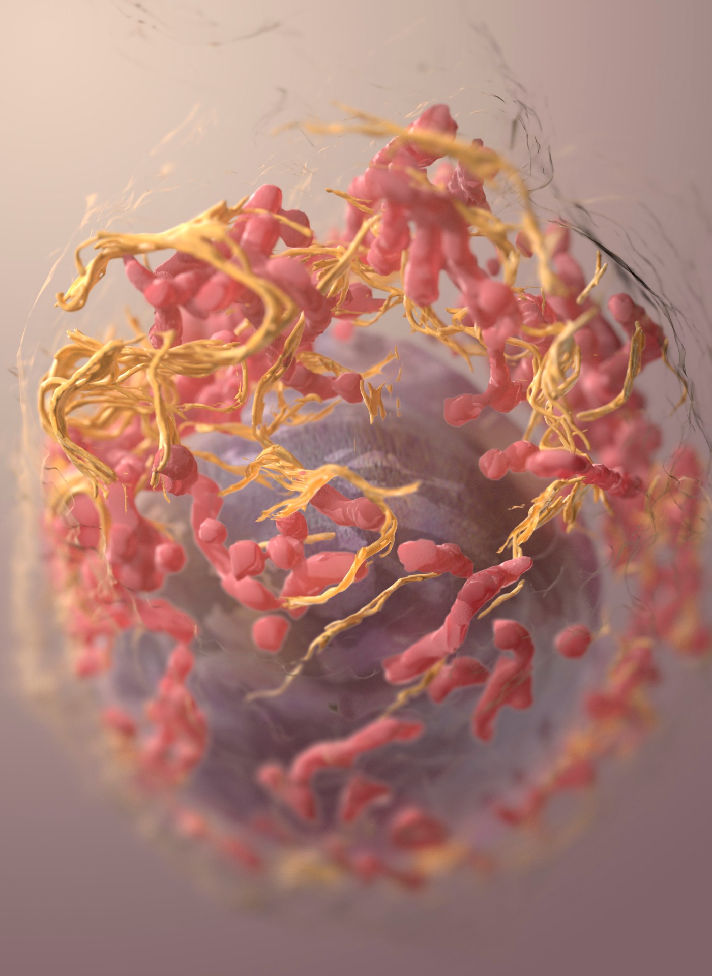
3D structure of a melanoma cell derived by ion-abrasion scanning electron microscopy. Sriram Subramaniam, National Cancer Institute, National Institutes of Health
