Reading time: 5 minutes
Sandhyaa Venkatachalam
Cervical cancer is jinxed in the world of oncology: caused by a high-risk HPV (Human papillomavirus) infection in the uterine cervix (lower part of the uterus/womb), it is one of the few cancers that can be gotten rid of, if detected early and through vaccination; yet as per the global cancer statistics 2020, remains a silent killer that dismally affects more than 600000 women globally, claiming over 300000 lives annually.
It develops at a very slow pace, usually beginning with precancerous lesions in the epithelium (cells lining the surface) of the cervix, termed as cervical intraepithelial neoplasia (CIN). Although challenging, tracking these initial changes can aid in timely treatment to nip it in the bud.
According to the World Health Organization, the standard method till date to diagnose cervical cancer is the Pap smear test, wherein cells from the cervix are scraped and examined under a microscope. If the cells seem suspicious, a biopsy may be performed. But this method has its limitations, because during the early stages, abnormal cells stealthily creep under a healthy layer of cells, at the junction between two epithelial layers of the cervix, rather than appearing on top of the surface. Hence, the scraped portion might not include the affected cells, thereby reducing the accuracy of the method.
The Circularly Polarized Light Scattering Technique:
A paper newly published in 2026 by Nishizawa N. and his team has garnered interest for proposing a modern technique based on circularly polarized light scattering (CiPLS). In order to fully comprehend the concept of CiPLS, it is essential to know what ‘polarized light’ means.
As explained by Manion GN & Stokkermans TJ, light is a form of electromagnetic wave that vibrates in all directions as it travels through space. Nevertheless, a polarized light stream can be created by filtering ordinary light through polarizers and wave plates, so that it waves only in one direction or a specific pattern. Likewise, in circular polarization, the field of light rotates in a circular motion as it moves forward (Figure 1).
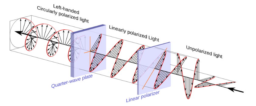
Figure 1: A quarter-wave plate transforms linear polarization into circular polarization and vice-versa.
When polarized light is shone at living tissues, it gets scattered when it encounters microscopic cellular structures such as nuclei, depending on their size and shape. And so, cancer cells typically having larger and more irregular nuclei than normal cells result in scattering that is markedly different from that of non-cancerous cells.
In a previous study published in 2024, Nishizawa N. reported that a critical advantage of using circularly polarized light is that it can maintain its polarization even whilst traversing through many layers of tissue, in contrast to linear polarized light which de-polarizes quickly.
CiPLS Experimental Design:
To test this idea, the researchers designed an experimental system which included artificial tissue samples, light sources (LEDs), polarizing optical components, and a special polarization-sensitive camera. Near-infrared light whose wavelength can penetrate up to 3 millimeters into the tissues without losing polarization, was preferred.
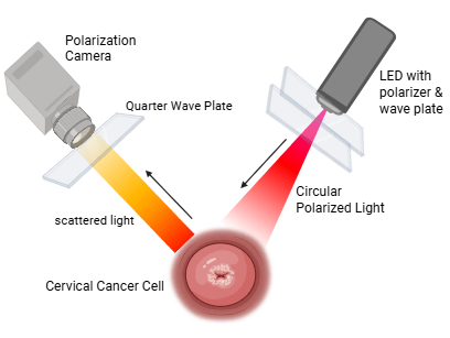
Instead of using tissues from real patients, they created a set-up that mimicked the layered structure found in cases of early cervical cancers. It consisted of a cancerous tissue layer sandwiched between two healthy layers (Figure 2). This design allowed them to thoroughly inspect how scattering changes depending on the depth and thickness of the cancerous layer.
Figure 2: Experimental set-up mimicking early-stage cervical cancer where the cancerous growth is hidden below a healthy layer.
Light was circularly polarized using a linear polarizer and a quarter-wave plate, after which it was directed onto the set-up at an optimal angle of 30° (angular measurements increase sensing accuracy). The light scattered in many directions due to interactions with the cell nuclei. The scattered light returning from tissues was captured by the polarization imaging camera after passing through a quarter-wave plate.
With the help of images generated, researchers deciphered a degree of circular polarization (DOCP) value, which indicates how much of the circular polarization signal is retained in the scattered light. Depolarization of polarized light due to multiple scattering depends on the ratio of the wavelength of light to the diameter of nuclei. Therefore, the research team could infer about the type of cells that were present within the layers, whether cancerous or non-cancerous.
Observations & Discussions:
It was observed that when the thickness of the upper healthy layer increased, i.e. the cancerous layer was buried deeper, the DOCP values noticeably changed. This implies that circularly polarized light scattering is useful not just to gain insights about the presence of abnormal tissues but also regarding the depth at which it is growing beneath the surface.
This work represents an important milestone towards developing a noninvasive diagnostic tool. Unlike the traditional biopsy method, this optical technique does not require tissue removal, staining, or biomarkers. It works in-situ (on-site) and in future, could potentially be integrated into medical instruments such as endoscopes. Such a device can reduce discomfort for patients, allowing doctors to effectively monitor any premature signs of cancer.
The study strongly emphasizes the need for further research before the technique can be implemented in clinical practices. Since researchers here have tested only on artificially layered tissues, the next step forward would be to test on actual samples obtained from patients. Other factors pertaining to pathological tissues that could influence the extent of scattering and subsequent polarization measurements should be taken into account such as cell density, cell membranes, cytoplasmic components, organelle variations eg. mitochondria, and detailing in the structures of epithelial layers. Needless to say, up-gradation in optical devices and imaging systems is indispensable to achieve uniform polarization and higher precision.
Header Image Source: Created by author in BioRender.com
Figure 1 source: https://commons.wikimedia.org/wiki/File:Circular.Polarization.Circularly.Polarized.Light_Circular.Polarizer_Creating.Left.Handed.Helix.View.svg
Figure 2 source: picture made in Microsoft Word
Edited by: Dr. Chiamaka Wisdom-Asotah
References:
- Nishizawa N, Ishikawa M, et al. Circularly polarized light scattering imaging of a cancerous layer creeping under a healthy layer for the diagnosis of early-stage cervical cancer. Journal of Biomedical Optics 31(2), 027002 (2026).
- Nishizawa N, et al. Depolarization diagrams for circularly polarized light scattering for biological particle monitoring. Journal of Biomedical Optics 29(7), 075001 (2024).
- Sung H, Ferlay J, et al. Global cancer statistics 2020: GLOBOCAN estimates of incidence and mortality worldwide for 36 cancers in 185 countries. CA: A Cancer Journal for Clinicians 71(3): 209-249 (2021).
- World Health Organization. Cervical cancer screening indicator metadata registry [Internet]. Geneva: WHO; cited 2026 Apr 12. Available from: https://www.who.int/data/gho/indicator-metadata-registry/imr-details/3240
- Manion GN, Stokkermans TJ. Polarization of light. In: StatPearls [Internet]. Treasure Island (FL): StatPearls Publishing; 2024 [cited 2026 Apr 12]. Available from: https://www.ncbi.nlm.nih.gov/books/NBK592424/

Leave a comment