
Reading time: 5 minutes
Madeline J. Morrisson
Ovarian Cancer
Ovarian cancer (OC) is the deadliest gynecological malignancy. High-grade serous ovarian carcinoma (HGSOC) is the most prevalent subtype of OC, with the majority of patients having late stage clinical detection. Like many types of cancer, HGSOC has both suppressive and protective immune mechanisms within the tumor microenvironment.
Advanced stage HGSOC has an increased number of regulatory T cells (Tregs). Tregs are a subtype of T cell that regulate, or suppress other immune cells1. T cells, along with other immune cells, have historically been categorized and defined by proteins on the cell surface or within the cell itself. Cell surface proteins allow T cells to interact with other immune cells, and proteins within the cell can control gene expression.
The vast majority of Tregs express the CD4 receptor, meaning they release signaling molecules known as cytokines to recruit other immune cells to a site of interest, which in the case of this paper, is a tumor microenvironment (TME).
Unlike other T cells, Tregs express an internal cell protein called FOXP3 (forkhead box P3), a transcription factor that regulates suppressive function, stability and expansion 2. Tregs act as the “brakes” within the immune system, and suppress the function of other immune cells via the release of cytokines. Because of this, they may be referred to in some cases as “anti-inflammatory” immune cells or immunosuppressive cells 1.
By leveraging sequencing data on samples from incidentally diagnosed patients, in “Immune evasion mechanisms in early-stage I high-grade serous ovarian carcinoma: insights into regulatory T cell dynamics” Milkulak et al found significant activation, infiltration, and differentiation of Tregs in early stage HGSOC 3.
Experimental Set up
Single cell RNA sequencing was performed on tumors and blood from two stage I HGSOC patients, who had not undergone chemotherapy. Diagnosis at stage I is rare, occurring in approximately 8.6% of those diagnosed with HGSOC 4.
Within the tumor samples, immune and non-immune cells were sequenced and analyzed separately. This is often done for samples that contain many cell types, to ensure that cells that only make up a small portion of a sample are not lost during sample preparation. A total of 12 populations of cells including immune cells, fibroblasts, and epithelial cells were identified. Focusing specifically at the immune cells, 15 different subtypes of CD4 T cells were found, many of which had an “activated” profile, meaning they are primed to multiply and respond to cancer. Understanding the interplay of these different cell types can lead to a greater understanding of how HGSOC progresses as well as generate possible targets for treatment.
Treg Heterogeneity in the Tumor Microenvironment
Within the CD4 T cells, a large degree of heterogeneity, or diversity, was observed between the two samples, Tregs were present in both. A total of 5 Treg subpopulations were identified: naive Tregs, immunosuppressive Tregs, cycling Tregs, cytotoxic Tregs, and ex-Tregs (Tregs with markers of instability). Notably, the immunosuppressive Tregs were found to highly express LAYN, a T cell receptor gene, that is associated with poor prognosis in OC.
ex-Tregs were defined and characterized by their loss of FOXP3 and by their pro-inflammatory characteristics. These cells have transcriptional instability, and as a result are not as effective at immune suppression. Pro-inflammatory signaling, derived from other cells within the TME, reprograms Tregs to ex-Tregs. In melanoma and head and neck squamous cell carcinoma, interferon gamma (IFN-γ) has been shown to drive the transition of Treg to ex-Treg. Increased expression of downstream targets of IFN-γ, that is, the other proteins and factors impacted by IFN-γ, within the ex-Treg was observed in HGSOC. Therefore, IFN-γ is a possible driver for Treg instability in stage I HGSOC.
Instability of Tregs leads to expression of CXCL13, a chemokine that recruits B cells and additional T cells to the TME. CXCL13 is correlated with patient survival, possibly because of how it drives the initial formation of tertiary lymphoid structures (TLS). TLS are clumps of immune cells that form near a tumor and coordinate the anti-tumor immune response 5.
Using a program called NicheNet, interactions of Tregs with other cell types were predicted. Broadly, Tregs are likely inhibiting DCs, CD8 T cells, and NK cells, aligning with their immunosuppressive function. Interactions between the Tregs and the cancer cells may be drivers of Treg activation. Tumor associated macrophages (TAMs), dendritic cells (DCs), and cytotoxic CD8 and NK cells were also found to be infiltrating the tumor. This aligns with what is typically observed within tumor tissue.
Conclusions and Next Steps
Findings from within this paper were confirmed using advanced stage HGSOC data from the Cancer Genome Atlas (TCGA), which showed similar expression patterns. This finding also supports the hypothesis that features of advanced stage tumors are already present in early-stage disease. This study is limited due to the small patient number (ie 2).
While additional work is still needed, Treg stability shows to be an exciting area of research within the ovarian cancer field. With this publication, we gain a greater understanding of ex-Tregs, which are driven to this phenotype by proinflammatory IFN-γ signaling, and express CXCL13 as a result. Increased CXCL13 may contribute to immune boosting in HGSOC patients. Whether these molecules or their associated pathways are viable for targeted treatment will need further investigation before movement into use in humans.
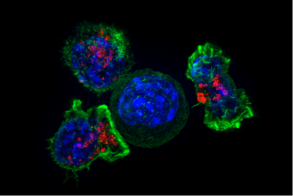
Header Image Caption and Source: Representative image of T cells (outer green stain) surrounding cancer cells (inner blue stain). Red stain marks granules, which contain chemicals that can kill undesirable cells. Retrieved 4/12/26: https://www.flickr.com/photos/nihgov/20673870162
Edited by Dolores Mruk
References
- Moncrieffe, H. (2023). Regulatory T Cells (Tregs). British Society for Immunology, Retrieved March 20, 2026, from https://www.immunology.org/public-information/bitesized-immunology/cells/regulatory-t-cells-tregs
- Thakur, S. (2021, June 25). FOXP3. Pathology Outlines. Retrieved March 20, 2026, from https://www.pathologyoutlines.com/topic/stainsfoxp3.html
- Mikulak, J., Terzoli, S., Marzano, P. et al. Immune evasion mechanisms in early-stage I high-grade serous ovarian carcinoma: insights into regulatory T cell dynamics. Cell Death Dis 16, 229 (2025). https://doi.org/10.1038/s41419-025-07557-5
- Matsuo, K., Machida, H., Matsuzaki, S., et al. (2020). Evolving population-based statistics for rare epithelial ovarian cancers. Gynecologic oncology, 157(1), 3–11. https://doi.org/10.1016/j.ygyno.2019.11.122
- Zhao, L., Jin, S., Wang, S. et al. Tertiary lymphoid structures in diseases: immune mechanisms and therapeutic advances. Sig Transduct Target Ther 9, 225 (2024). https://doi.org/10.1038/s41392-024-01947-5


Leave a comment