
Reading time: 5 minutes
Emily Chan
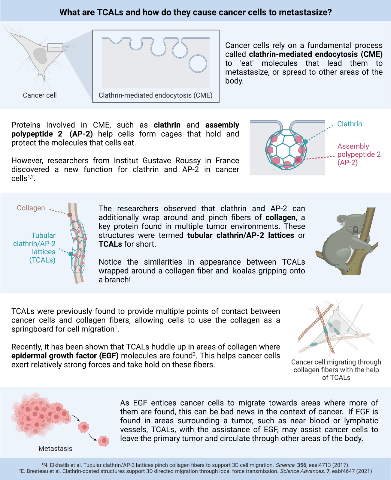
During a holiday weekend, I had the chance to see one of the most beloved animals at my local zoo: the koala. Contrary to their adorable exterior, koalas boast strong arms and legs that hook around tree branches, allowing them to scale large eucalyptus trees. Recently, researchers at Institut Gustave Roussy in France discovered that cancer cells use their own type of koala-like structures to “grip” around networks of collagen fibers embedded in tumor environments. The researchers showed that cells that possess these structures, known as tubular clathrin/AP-2 lattices (TCALs), migrate more efficiently than cells without TCALs (see Figure 1). This is bad news for cancer cells that can exploit this trait to metastasize, or migrate and form tumors in other areas of the body. So, how does this occur?
A recent study, published in Science Advances by the founders of TCALs, explored this question by first setting up a platform to study TCAL formation during cancer cell migration. Cancer cells were placed on collagen fibers coated with epidermal growth factor (EGF), which has been shown to bind to its receptor, epidermal growth factor receptor (EGFR) at the cell surface and activate signaling pathways that prompt cells to migrate. After verifying that the cancer cells could sense EGF on collagen, EGF, EGFR, and TCALs were labeled so that they could be viewed under the microscope. As expected, EGFR clustered more frequently in areas where the collagen was coated with EGF than areas without EGF. Similarly, TCALs were observed to form in greater quantities and cluster more quickly in areas where EGF was found on collagen, and this was dependent on the presence of EGFR. However, this process was not dependent on signaling pathways prompted by activating EGFR that lead to uncontrolled cell division and survival, hinting that EGF-induced clustering of TCALs and EGFR on collagen acts as a separate mechanism that cancer cells use to function in tumor environments.
To see whether this function is preserved during the initial stage of cancer cell migration, known as cell spreading, the researchers probed TCAL and EGFR formation as cancer cells stretched their cell membranes onto collagen. Cell membranes elongated on bare collagen fibers, but elongated even more on EGF-coated collagen fibers in the presence of TCALs and EGFR. The researchers then used a microscopy technique that allowed them to measure the amount of force that cells create as they spread on collagen. Cancer cells exert more force when they spread on EGF-coated collagen than bare collagen fibers. While this occured, the researchers further noticed that collagen fibers located at the top side of cells transitioned from straight and thin bundles to spherical clumps, indicating that cells remodeled the collagen in this area. The collagen fibers that were coated with EGF were not only remodeled in greater quantities by the presence of TCALs and EGFR, but faster than bare collagen fibers. From this set of experiments, the researchers concluded that in addition to their clustering abilities, TCALs help cancer cells elongate and exert more force on EGF-coated collagen fibers than cells on bare collagen fibers. These forces are created at specific areas of the membrane to help cells remodel their environment as they begin to migrate. Similar to the previous findings, these properties were not dependent on EGFR activation, strengthening the characterization of TCALs as unique koala-like structures that cancer cells use to efficiently grip onto and probe tumor environments.
The last piece of the puzzle was to determine whether these koala-like grips assist cancer cells to metastasize, or migrate in tumor environments. The researchers initially observed cancer cells migrating on collagen coated with EGF that was equally distributed everywhere. TCALs didn’t seem to help cancer cells move as they migrated at the same rate on EGF-coated and bare collagen fibers. The researchers then changed their setup to account for a key cause of metastasis, known as chemotaxis, in which molecules such as EGF orient and entice cancer cells to move to locations where more EGF is found. Like how a koala moves to another tree to indulge in more leaves, the researchers observed cells migrating away from bare collagen towards EGF-coated collagen fibers using their TCALs/koala-like grips. This is a dangerous finding in the context of cancer. If EGF is found in areas surrounding a tumor, such as near blood or lymphatic vessels, TCALs, with the assistance of EGF, may assist cancer cells to leave the primary tumor and circulate through other areas of the body.
Future studies to probe the function of TCALs during cell migration need to be performed within environments that incorporate multiple aspects of a tumor. For example, it is unknown whether non-cancerous cells, which are also key components of tumors, similarly exploit TCALs to migrate. The Institut Gustave Roussy researchers took the lead on attempting to answer this question by uncovering that fibroblasts, which are non-cancerous cells that produce collagen, create membranous tracks on top of collagen fibers that guide cancer cell migration in tumor environments. While there is some evidence that cancer cells use TCALs to migrate on these fibroblast-produced tracks, whether fibroblasts themselves possess TCALs is an open question. Targeted treatments towards TCALs could be a possibility in the future if new studies reveal a substantial difference in how cancerous and non-cancerous cells use TCALs to migrate. That being said, this study illuminated the importance of TCALs as structures that congregate in certain areas of a tumor where there are cancer-promoting molecules like EGF which help cancer cells create sufficient forces to migrate towards these locations.
Edited by Payal Yokota
Photo by Elmer Cañas on Unsplash
Primary works cited:
F. Baschieri, A. Illand, J. Barbazan, O. Zajac, C. Henon, D. Loew, F. Dingli, D. M. Vignjevic, S. Lévêque-Fort, G. Montagnac, Fibroblasts generate topographical cues that steer cancer cell migration. bioRxiv. (2022). https://doi.org/10.1101/2022.09.06.506801
E. Bresteau, N. Elkhatib, F. Baschieri, K. Bellec, M. Guérin, G. Montagnac, Clathrin-coated structures support 3D directed migration through local force transmission. Science Advances. 7, eabf4647 (2021). https://doi.org/10.1126/sciadv.abf4647
N. Elkhatib, E. Bresteau, F. Baschieri, A. L. Rioja, G. van Niel, S. Vassilopoulos, G. Montagnac, Tubular clathrin/AP-2 lattices pinch collagen fibers to support 3D cell migration. Science. 356, eaal4713 (2017). https://doi.org/10.1126/science.aal4713

Leave a comment