Reading time: 4 minutes
Nisitha Sengottuvel
Cancer’s got nerve. We began discussing this earlier in the month with a blog post by Katelyn Fleishem. While neuronal activity has been implicated in the progression of tumor progression in prostate cancer, stomach cancer, colon cancers among others, the mechanisms of what role these neurons play are just now beginning to be worked out. So far, much of what is known about the mechanism has to do with signaling from neurotransmitters and their downstream effects. Neurotransmitters are molecules released at the regions where nerves make connections, used to send messages throughout our body. They have been shown to promote important processes of cancer such as triggering Wnt signalling, a well known signaling pathway involved in numerous pro-cancer processes including promoting increased cell divisions among cancer cells. Neurons are known for their electrical stimulation as a relay system of communication in our body. The work regarding neurons and cancer has just now begun to dive into the mechanistic implications of electrical stimuli from neurons leading to cancer progression.
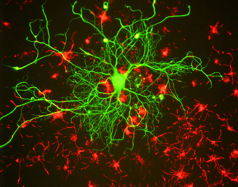
Recently, Venkatesh et al. published a paper about electrical and synaptic integration of glioma into neural circuits. Gliomas are a type of deadly brain cancer. In fact, these cancers are the leading cause of death from tumors arising from the central nervous system. According to Mesfin and Al-Dahir, there are 80,000 new primary brain tumors diagnosed every year and about 25% of these cases are gliomas. This group had previously found that gliomas enrich genes that make the cancer cells look like neuronal synapses. Synapses are the junctions between two neurons where communication occurs, either electrical or chemical. The fact that the glioma cells make themselves look more like synapses falls right in line with the thinking that cancerous cells tend to adopt their gene expression to match different components of the host’s body in order to fit in and take advantage of our body’s resources to grow.
Since these cells start “looking” like neurons by increasing genes that make them look like a synapse, this group next began trying to understand whether the neurons and glioma cells made functional synapses. They wanted to make sure that the neurons and glioma cells were able to “talk” to each other by passing electrical messages between themselves. They used a fluorescent label to identify glioma cells and placed these cells in the hippocampus (a region of the brain with high levels of electrical activity). After they let these glioma cells grow, they took slices of tissue from the hippocampus that included cancer cells from the glioma as well as neurons. They stimulated axons of the neurons in this slice and measured the voltages in the glioma cells to see if when the neurons are stimulated, there was an electrical response in the tumor cells as well. To confirm that the electrical activity they read in the glioma cells were due to communication with neurons, they used an inhibitor called tetrodotoxin which blocks voltage gated sodium channels. These sodium channels are important in sending electrical signals between neurons. When these channels are blocked, they saw that the voltage recordings stopped showing stimulation in the cancer cells. This suggested that the glioma cells not only adopt gene expressions that look like a neuron, but also functions like one by being able to communicate with other neurons.
In preclinical mouse models of glioma, previous studies had found that there was hyperexcitability and seizures induced in these tumor models. Venkatesh et al. also found that the glioma cells, on top of genetically and functionally mimicking a neuron, cause neighboring neurons to become highly excitable. In order to measure this, they took three (human) patients who had high grade gliomas and used electrocorticography to measure the electrical activity in the brain. They measured this electrical activity for three minutes while the patients were awake in a resting state and found that in regions with tumor, there appeared to be much higher electrical activity than in “healthy appearing” brain regions.
Neuronal activity is becoming a hot topic in cancer research, not only for brain cancers, but for many different types of cancers. Now that the field of oncology research is beginning to appreciate the importance of nerves in the tumor microenvironment, the next step is to figure out what these nerves are doing there. This study by Venkatesh et al., begins uncovering the electrical side of the mechanistic value that neurons bring in tumor progression. Next, it would be interesting to understand if tumor cells coming from other cancers (outside of the brain) are also able to induce hyperexcitability of nearby invading neurons. Taking these findings and applying them to other cancers will be the key to increasing the breadth of where these findings will take the field of oncology.
Edited by Kedar Puvar
Work Discussed
Venkatesh, Humsa S., et al. “Electrical and Synaptic Integration of Glioma into Neural Circuits.” Nature, vol. 573, no. 7775, 2019, pp. 539–545., doi:10.1038/s41586-019-1563-y.
Image Credits

Leave a comment