Reading time: 4 minutes
Varshit Dusad
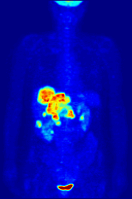
 Studying the growth of the tumor is very important to devise an effective treatment against cancer. Often cancer tissues are buried deep inside the body and we need diagnostic methods that can tell us about their progression accurately without harming the body while not losing the accuracy. Therefore, invasive surgery is not an option. A very common method is isotopic labeling. This relies on a specific atom’s radioactivity, or tendency to release extra energy in the form of radiation. Many common elements including carbon and nitrogen have radioactive isotopes; the stable nitrogen in the air is only one version of nitrogen, while others can be unstable and release radioactive energy. Apart from their scarcity and radioactivity, they have identical chemical properties to their non-radioactive counterparts. These radioactive isotopes can be substituted into larger chemicals in place of normal atoms and, because they release radiation predictably, can be used as a label for whatever chemical they’ve been incorporated into. One such labeled chemicals 18F Fludeoxyglucose (or 18FDG) which resembles the natural sugar glucose in its chemical structure with a small change. It possesses radiolabelled fluorine instead of a hydroxyl group in natural glucose. It is very commonly used in medical imaging techniques, including PET scans.
The idea behind using radiolabelled 18FDG is simple. Instead of normal glucose, the patients are asked to consume 18FDG. The benefit is that unlike regular glucose the movement of 18 FDG inside the human body can be monitored using PET. Here at OncoBites, we have previously talked about the important role of metabolism in understanding cancer and developing anti-cancer treatments. We have discussed how cancer cells have a higher rate of consumption of glucose and other nutrients compared to healthy cells. Due to its similarity to glucose, 18FDG is also consumed more by cancer cells than normal cells. However, the little change of possessing radiolabelled fluorine and instead of hydroxyl group prevents it from being broken down inside the human body the way normal glucose is. Thus, once consumed it stays put in the tissues and because cancer tissues have a higher tendency to absorb it, it accumulates inside tumors. Due to this accumulation, the radioactive-emission is higher from these tissues compared to the rest of the body. Thus, it is easily monitored from outside the body as tumors show up as bright spots on the scan.
However, this is only so simple in an ideal world. In reality, the uptake of 18FDG across the tumor tissue is not consistent. A common observation is that tumor cells at the outer edge of the tissue take up more 18FDG than the cells at the core of tumor tissue. For a long time, it was assumed that this was probably because it’s easier for the cells on the outer edge to access the 18FDG from the bloodstream. However, a recent study found that this assumption was not true, and the lack of uniform 18FDG uptake has a deeper connection to cancer metabolism than previously understood. The authors of this study investigated different plausible hypotheses to explain this observation. Out of which, they rejected hypoxia i.e. lack of oxygen in an environment which is more prevalent inside core tumor cells as a possible reason. This was because lack of oxygen should ideally increase the 18FDG uptake and not decrease it. Similarly, the growth rate between the core and periphery cells was also rejected due to inconclusive evidence. Eventually, based on experimental data, the scientists concluded that it was actually the presence of higher amount of lactate in core tumor cells which resulted in poor uptake of 18 FDG. The presence of lactate, as well as low pH (increased acidity), actively suppressed the transport of 18FDG inside of core cancer cells. The core cells possess a higher amount of lactate, possibly due to their hypoxic environment which promotes anaerobic respiration that produces lactate.
This study reveals another interesting and important role the metabolism plays in cancer biology. In a typical clinical setting, the low uptake of 18FDG will be considered as a sign of tumor regression or even absence of cancer. However, these latest results prove that the obvious results coming from PET can be highly deceiving. We have discussed before at OncoBites that increased lactate metabolism (also called Warburg effect) is an integral part of cancer physiology and has been used as a basis for developing anti-cancer medicine. There is still an immense need for improving the resolution and accuracy of our cancer diagnostics tools.
Literature Cited:
Turkcan, S., Kiru, L., Naczynski, D. J., Sasportas, L. S., & Pratx, G. (2019). Lactic Acid Accumulation in the Tumor Microenvironment Suppresses (18)F-FDG Uptake. Cancer Res, 79(2), 410-419. doi: 10.1158/0008-5472.CAN-17-0492
