Reading time: 5 minutes
Jessica Campbell
Background
Arguably, the most dangerous aspect of cancer progression is when the cancer cells leave the original primary tumor and migrate away to find a new home elsewhere in the body. A single primary tumor is much easier to treat than multiple tumors that may be in multiple locations in the body. This is why early detection and diagnosis of a primary tumor by screening processes like mammograms and smear tests is essential. Unfortunately, early detection is not always possible, and understanding how and why cancer cells migrate (metastasize) may help provide new therapeutic avenues for doctors to target.
Primary tumors are formed when healthy cells collect mutations in their DNA, causing these cells to make too many copies of themselves. At the start of this process, tumors are not necessarily cancers and are often just benign lumps that may never progress or cause harm. It is only when these tumor cells start invading the surrounding tissue that we call them cancer, and these cells become malignant. To begin the invasion process, cells will switch on genes and create particular proteins to allow the cells to become motile. This motility is driven by the cell’s skeleton (cytoskeleton), which will change shape and physically push on the outer cell layer (cell membrane) to propel the cell forward, dragging its rear behind it. Cells in the body must travel through a dense 3D network of criss crossing fibres, called the extracellular matrix (ECM), before they find a new destination to settle down and make secondary tumours. During their journey, cancer cells will interact with healthy cells, including immune cells, blood vessel cells, and a specialized type of cell called cancer-associated fibroblast (CAF). Controversially some of these healthy cells, CAFs in particular, have been shown to promote tumor cell growth and migration, but how exactly do they do this?
New research
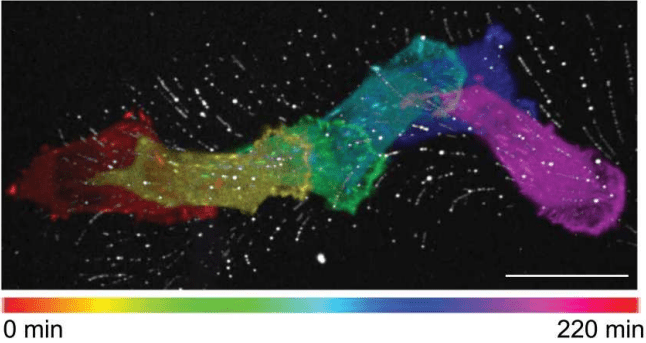
Recently, scientists at the Universite Paris-Saclay have used a number of elegant experiments to investigate the relationship between CAFs and breast cancer cells (see further reading article 3). Using microscopy techniques, the authors saw that as the CAFs migrated, they laid out behind them a ‘network of membranous material’, like a trail of breadcrumbs (Figure 1). This was true when CAFs migrated on a 2D glass dish and within a 3D ECM-like network. The authors termed this material ‘tracks’ and found that the tracks remained for days after the CAFs had migrated away. The branches of the tracks were at similar angles, pointing in the direction of the CAF cell migration and were bound tightly to the 2D glass surface.

These tracks were not made from an ECM protein such as collagen, as the scientists may have expected them to be, but were in fact made from the CAF cell membrane – the CAF cell was leaving little pieces of itself behind.
In order to study how these tracks affected cancer cells, the authors allowed the CAFs to lay down their tracks on a 2D surface and then removed the CAFs, and replaced them with breast cancer cells. They found that the breast cancer cells preferentially stuck to the surfaces coated with tracks and migrated along these tracks in the direction the CAF originally did (Figure 2). As the cancer cell migrated, some of the tracks disappeared beneath it, and using super-resolution microscopy, the authors found that the cancer cell internalized some of the tracks. They hypothesized the tracks form part of a communication system between CAFs and cancer cells, but what happened to the tracks once they became internalized was unclear. Further experiments were conducted to investigate the proteins required for the cancer cell-track interaction and the mechanism of track internalization. For more information and details, please look at the original paper. There are some lovely movies of the cells migrating around!

Conclusion
This study suggests that one mechanism for cancer cells to migrate away from their primary tumour to form secondary tumours is to follow these tracks, however, many questions remain. What happens to the sections of tracks that have become internalized by the cancer cells? What protein signaling pathways are activated within the cell? How does the cytoskeleton orient itself in the correct direction? And importantly, are there any therapeutic ways to prevent this from happening in the body? Future work will undoubtedly aim to answer these questions.
Edited by Rachel Cherney
Further reading
1. For a general understanding of how cancers develop see ‘Hallmarks of cancer: the next generation’
Hanahan D, Weinberg RA. Hallmarks of cancer: the next generation. Cell. 2011 Mar 4;144(5):646-74. doi: 10.1016/j.cell.2011.02.013. PMID: 21376230.
2. To read more detail about cancer associated fibroblasts see ‘A framework for advancing our understanding of cancer-associated fibroblasts’
Sahai E, Astsaturov I, Cukierman E, DeNardo DG, Egeblad M, Evans RM, Fearon D, Greten FR, Hingorani SR, Hunter T, Hynes RO, Jain RK, Janowitz T, Jorgensen C, Kimmelman AC, Kolonin MG, Maki RG, Powers RS, Puré E, Ramirez DC, Scherz-Shouval R, Sherman MH, Stewart S, Tlsty TD, Tuveson DA, Watt FM, Weaver V, Weeraratna AT, Werb Z. A framework for advancing our understanding of cancer-associated fibroblasts. Nat Rev Cancer. 2020 Mar;20(3):174-186. doi: 10.1038/s41568-019-0238-1. Epub 2020 Jan 24. PMID: 31980749; PMCID: PMC7046529.
3. To read the full paper discussed in this article see ‘Fibroblasts generate topographical cues that steer cancer cell migration’- don’t forget to watch the movies!
Baschieri F, Illand A, Barbazan J, Zajac O, Henon C, Loew D, Dingli F, Vignjevic DM, Lévêque-Fort S, Montagnac G. Fibroblasts generate topographical cues that steer cancer cell migration. Sci Adv. 2023 Aug 18;9(33):eade2120. doi: 10.1126/sciadv.ade2120. Epub 2023 Aug 16. PMID: 37585527; PMCID: PMC10431708.

Leave a comment