
Reading time: 4 minutes
Jessica Desamero
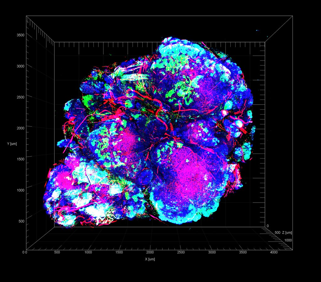
With cancer, the environment that surrounds a tumor, or the tumor microenvironment (TME), can be just as important as the tumor itself in terms of cancer progression and drug resistance. How can this be? First, let’s take a look at what the TME is composed of.
In a tumor microenvironment, there are, of course, cancer cells. Next, there are many blood vessels and other types of cells within the vicinity. These types of cells include those of the immune system, fibroblasts, which are cells that produce fibers like collagen, and cells that line the interior surfaces of blood vessels, called endothelial cells. Then there are additional components, including the extracellular matrix, which is important for structural support, and molecules involved in cellular communication.
Cancer cells interact with their environment through complex networks of signals. The extent of these interactions and environmental conditions can influence whether or not these cancer cells continue to proliferate and spread through other areas of the body.
Nearly all solid tumors display hypoxia, or a lack of oxygen in the tumor and surrounding tissue. As cancer cells rapidly multiply, they become too numerous to be supported by surrounding blood vessels. Consequently, the availability of overall oxygen decreases, leading to regions with low oxygen levels called hypoxic regions. Current anticancer strategies mainly target cancer cells around blood vessels, meaning tumor cells found in hypoxic regions that adapt to the lack of oxygen can proliferate and become more drug-resistant.
Some of these hypoxic region adaptations involve changes in the tumor microenvironment itself. For example, due to the increased need for oxygen, tumor cells develop new leaky blood vessels. Additionally, cellular respiration without oxygen leads to accumulation of the byproduct, lactic acid, thereby increasing the overall acidity of the microenvironment. This acidity can inhibit processes of certain leukocytes, which are immune cells that help protect the body from foreign invaders and infectious agents, alter genetics by promoting the occurrence of mutations, decrease the amount of chemotherapeutic drug in tumor cells, and enable cancer cells to develop resistance to oxidative stress-induced apoptosis, or programmed cell death. Moreover, stress from the lack of oxygen can suppress the general immune response.
Another set of adaptations involve a transcription factor called hypoxia-induced factor 1α, or HIF-1α. Transcription factors are proteins that can control the rate at which DNA is copied into RNA, and they can regulate when genes are expressed. As oxygen levels decrease, expression levels of the HIF-1α gene increase, making the HIF-1α protein more active. The HIF-1α protein then triggers signaling events that activate proteins that help transport drugs out of cells, inhibit apoptosis, overcome DNA damage, maintain cellular respiration efficiency, and enhance the development of cancer cells’ multidrug resistance. In addition, p53 is a protein that controls cell division and thus acts as a tumor suppressor. HIF-1α can directly reduce expression levels of p53, further preventing apoptosis and promoting cell proliferation.
To combat this increased drug resistance, multiple strategies to target hypoxic tumor cells have been proposed:
- Create drugs that are initially inactive but become active upon recognizing a particular substance found specifically in tumors completely lacking oxygen.
- Target and regulate HIF-1α. Drugs can be destined to trigger activation of HIF-1α degradation pathways or inhibit the expression of HIF-1α in the first place.
- Eliminate reactive oxygen species (ROS) resistance in tumor cells. ROS are overproduced in tumor cells. At high amounts, ROS can cause severe damage to biomolecules, which would normally cause cell death. Cancer cells, however, can survive this due to the activation of a defense system that prevents too much ROS from being produced and activates cell survival proteins. If the components of this system can be eliminated, accumulation of ROS could kill the tumor cells.
Hypoxia is one factor that continues to complicate the advancement of targeted therapies. It can render chemotherapy drugs and the immune system ineffective against tumor cells. But understanding more about cell death and the detailed impact of the hypoxic microenvironment on various cellular processes would greatly aid in developing more successful, longer-lasting cancer treatments.
Edited by Michael Marand
Work Cited
Jing, X., Yang, F., Shao, C. et al. Role of hypoxia in cancer therapy by regulating the tumor microenvironment. Mol Cancer 18, 157 (2019). https://doi.org/10.1186/s12943-019-1089-9
Image Credit

Leave a comment