
Reading time: 5 minutes
Garima Khanna

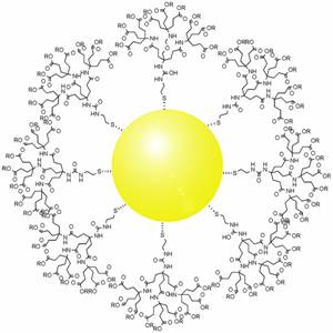
Gold nanoparticles (GNPs) possess exceptional characteristics, including high surface area to volume ratio, easy synthesis, surface chemistry, multi functionalization, stable nature and surface plasmon resonance. Because of these unique properties, they are emerging as a powerful tool for early tumor diagnosis and chemotherapeutic drug delivery targeted to the tumor site. Both radiation and chemotherapy, the two most common cancer therapies, are based on killing cancer cells. However, this killing is not selective and causes enormous damage to the surrounding healthy tissue. Gold nanoparticles offer the possibility of treatment which overcomes the limitations of current cancer therapies.
Enhanced Permeation and Retention Effect
The leaky blood vessels in tumor tissue may make it easier for small materials, such as gold nanoparticles, to move out of the bloodstream and into the tumor. This phenomenon, which is a highly debated topic in cancer biology, is known as the enhanced permeation and retention (EPR) effect. Nanoparticles accumulating in tumors through the EPR effect is known as passive targeting, and is rarely sufficient for treating human tumors. However, tumors can also be targeted through biomarkers, which are molecules specific to tumor cells; this process is known as active targeting. Active targeting uses molecules like monoclonal antibodies, aptamers, peptides and vitamins that are attached to the surface of nanoparticles, which interact with molecules on cancer cells; this process then brings the nanoparticle into the cell to release the drug. Active targeting is therefore a more powerful tool for anticancer therapies. .
Uses of Gold Nanoparticles
Traditional drug delivery disseminates the drug throughout the whole body, with only a fraction of the drug reaching the tumor site. GNPs have attracted researchers as drug carriers due to their ability to be prepared in a wide range of sizes, from as low as 1 nm to 150 nm, which gives us very precise control over their properties. GNPs are covered with a negative charge, allowing for modification with the addition of various biomolecules. Methotrexate, for example, is more toxic to numerous tumor cell lines when attached to gold nanoparticles than when it is given alone.
Photothermal therapy uses light to overheat small areas (hyperthermia), which causes cellular structures to break down,ultimately leading to cell death. Because gold nanoparticles come in many shapes and sizes, they can be used to absorb and reflect a wide range of light wavelengths (from UV to IR), and are more potent than conventional dyes.
Aurolase®, developed by Nanospectra, are silica-gold nanoshells coated with a biocompatible polymer (polyethylene glycol (PEG)) and designed to heat and kill solid tumors. The absorption of light leads to an increase in the local temperature, destroys solid tumors.
Photoimaging uses functionalized GNPs injected into the tumor site which specifically bind to cancer cells and tag them for surgical removal. This technique can help in detection of early stage tumors and guide the surgeons for precise treatment. Magnetic Resonance Imaging (MRIs) and Computed Tomography (CT) scans are limited and can only evaluate a tumor size above several millimeters. Functionalized gold nanoparticles, when injected into the tumor, bind to cancer cells and scatter light to help differentiate between tumor and healthy cells.
Due to their bioinertness and the ability to provide an efficient resolution, gold nanoparticles are considered the best agents for photoimaging in cancer therapeutics.
Despite several studies, there is still a need for affordable gold nanoparticle-based systems that improve the diagnosis and treatment of cancer at early stages, even though they possess various winsome characteristics. Certain limitations like inefficient biodistribution, toxicity and size propose a question for more validated data to further their use. A great deal of research is called for the development of efficient gold nanoparticles for therapeutic use. (Singh et al., 2018)
Edited by Sara Musetti
Works Discussed:
Singh, P., Pandit, S., Mokkapati, V. R. S. S., Garg, A., Ravikumar, V., & Mijakovic, I. (2018). Gold Nanoparticles in Diagnostics and Therapeutics for Human Cancer. International Journal of Molecular Sciences, 19(7). https://doi.org/10.3390/ijms19071979
Image Credits
Gold Nanoparticles in Cell Cytosol, NIH Image Gallery
Figure 1: Gold_Nanoparticles.jpg (300×300). (n.d.). Retrieved October 8, 2019, from Wikimedia
